Shocking! India’s first reported case of intrahepatic pregnancy: Foetus found growing in woman’s liver
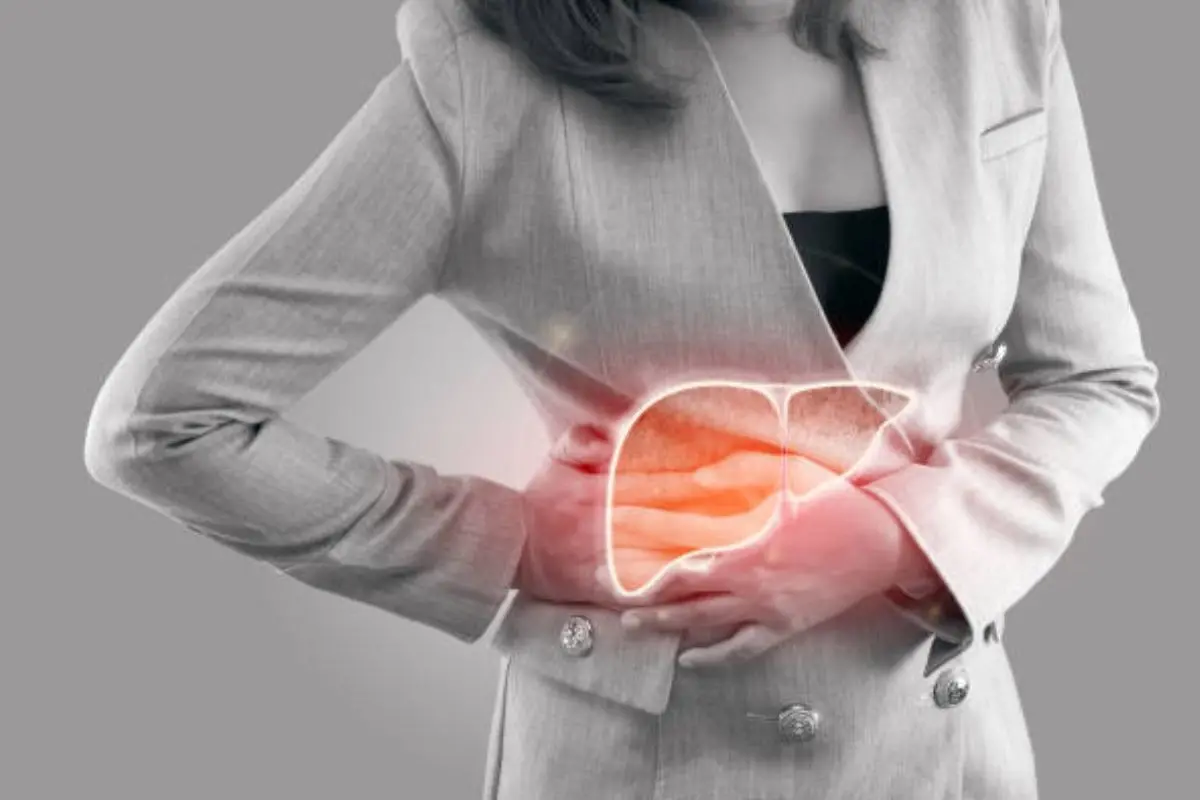
Uttar Pradesh: A 30-year-old woman from Bulandshahr was found to be 12 weeks pregnant, not in her uterus, but in her liver. The rare medical condition, known as intrahepatic ectopic pregnancy, is believed to be the first known case ever reported in India.
The woman had been experiencing continuous abdominal pain and vomiting for several weeks. When initial ultrasounds failed to find the reason for her symptoms, she was referred for an MRI of the abdomen.
The MRI was conducted at a private imaging centre in Meerut under the supervision of Dr. KK Gupta, a senior radiologist with decades of experience. “When I saw the scan, I could not believe my eyes. The foetus was embedded in the right lobe of the liver, and there were clear cardiac pulsations. I have never seen such a case in my career, and according to available data, this might be India’s first intrahepatic ectopic pregnancy," Dr Gupta explained to News18.
Unlike a routine ultrasound, the MRI provided high-resolution, cross-sectional images of the abdominal organs. According to Dr. Gupta, the scan showed a well-formed gestational sac located within the right lobe of the liver. The foetus was estimated to be around 12 weeks in gestational age, and what shocked the team even more was the detection of active cardiac activity, indicating the foetus was alive.
Further, to ensure the accuracy of the diagnosis, repeated imaging was done. Dr. Gupta said he initially considered the possibility of a technical error or an imaging artefact, but the repeated scans, taken from multiple angles, confirmed the presence of a live foetus embedded in the liver tissue.
Dr. Gupta also noted that the gestational sac appeared deeply implanted into the liver’s parenchymal tissue, with hepatic blood vessels supplying nutrients, further confirming that they were dealing with an extremely rare, high-risk pregnancy.
What is an ectopic pregnancy?
In a typical pregnancy, the sperm and the egg meet in the fallopian tube, after which the fertilised egg travels to the uterus and implants itself in the uterine wall to grow. However, when the fertilised egg implants and begins to grow outside the uterus, such as in the fallopian tube, cervix, or even within the abdominal cavity, this condition is known as an ectopic pregnancy.
How and why does it happen?
Though the exact cause of ectopic pregnancy isn't clear yet, one possible reason is damage or infection in the fallopian tube. Inside these tubes are fine, hair-like structures called cilia, which help move the egg toward the uterus. If an infection affects these cilia, their movement may be impaired, preventing the fertilised egg from reaching the uterus. As a result, the embryo may start growing inside the fallopian tube itself.
Since the fallopian tube is a narrow structure, it can support the growth of an embryo only up to about six weeks. Beyond that, the expanding pregnancy may rupture the tube, causing internal bleeding, a potentially life-threatening situation.
An ultrasound scan is usually performed within the first six weeks to confirm the pregnancy is developing inside the uterus.
Diagnosis and treatment options
If detected early, ectopic pregnancies can be treated with medication. However, this is only possible under specific conditions: the person must be in stable health, beta hCG levels should be below 3000 mIU/mL, the gestational sac should be smaller than 3.5 cm, and the heartbeat of the foetus should not start. After medication is administered, beta-hCG levels are closely monitored to ensure they return to their normal state. Follow-up is critical during this period.
If medication is not suitable or fails to work, it is necessary to have a surgical intervention. In emergency situations, especially if the tube has ruptured and severe internal bleeding occurs, immediate surgery is required. Laparoscopic surgery is often preferred, but in some cases, the affected fallopian tube might have to be removed entirely. If the embryo is located in the ampullary portion (the mid-section of the tube), a method called milking may be used to gently push it out. Alternatively, a small incision may be made in the tube to extract the embryo.